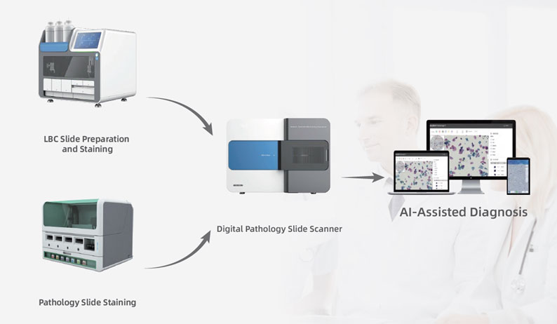
We offer small-to-large routine pathology laboratories high-quality and high-speed digital scanning solution, from low-to-high throughput applications. The digital pathology slide scanners are designed to meet diagnostic requirements, and guarantee constant color fidelity and high resolution histological and cytological samples, so that the image can be safely used to create a diagnosis. Powered by unique scanner software, the digital pathology slide scanners can achieve one-touch, fast and high-quality slide scanning to meet the needs of routine pathology laboratories.
Multiple Options:
12 slides, 120 slides, and 600 slides formats available.
Automated:
ONE click to achieve fully automated scanning operation.
Digital Image:
various file formats for case discussions, teleconsultation and education.
High Precision:
Multilayer fusion imaging technology (up to 15 layers).
High-quality:
Automated self-calibration during scanning for consistent image quality.
AI - assisted cervical cancer screening system
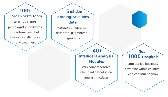
The AI assisted system offers comprehensive slide analysis for cervical cancer screening, with performance listed below, it can highly speed up the WHO’s objective to eradicate cervical cancer by 2030 via reducing pathologist’s workload and enhance the diagnosis efficiency.
Sensitivity: 5%
Specificity: 70%
Analysis: < 1 minute
Sample Evaluation
Automatically evaluate and analyze the effective cell quantity and quality to ensure sample effectiveness.
High-risk Area Mark
Intelligently mark the suspicious area, such as the section contains positive cells or microbial infections, for pathologist alert and double confirmation.
AI Recommendations
The system also generates AI recommendations for whole-slide images to support pathologists in making accurate diagnoses.
Excellent Assistant
Pathologists can quickly and efficiently screen large amounts of samples, identifying negative cases with the help of AI-assisted analysis.
High Compatibility
Compatible with a variety of scanner models
Unique Compress Technology
Lossless compress technology to maximize storage pace
Convenient and Versatile
Multiple access. Fast retrieval to facilitate tele-training Intelligent annotation tools available
Quality Control
API supports pathological info system ensures quality control of whole process
Objectivity and Stability
Quantitative analysis of whole-slide images to avoid subjectivity
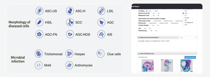
According to the requirements of TBS diagnostic criteria, a variety of diseased cell morphology and microbial infection types can be identified:
Instant Result: <2 mins. to get diagnostic report, hard copy available
Highly effective: AI-flagged positive samples categorized automatically according to TBS, , NLNM; ASCUS; LS