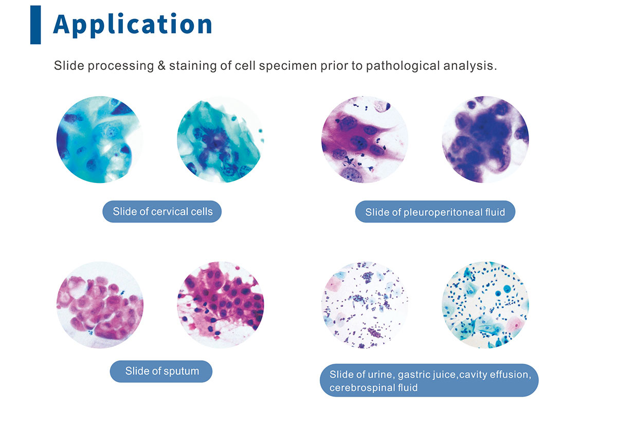
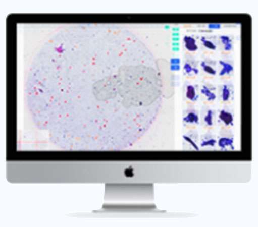
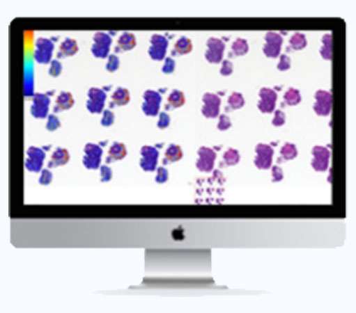
Morphology & Cytology
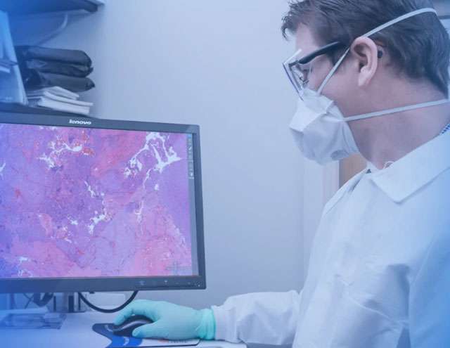
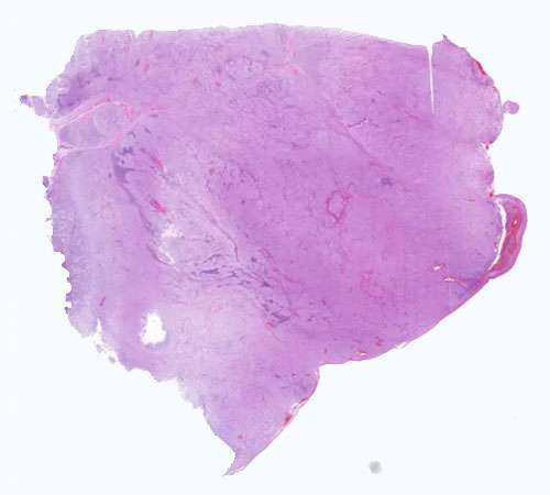
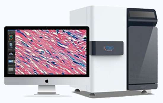
Digital Pathology Slide Scanner

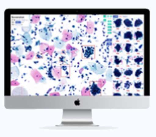
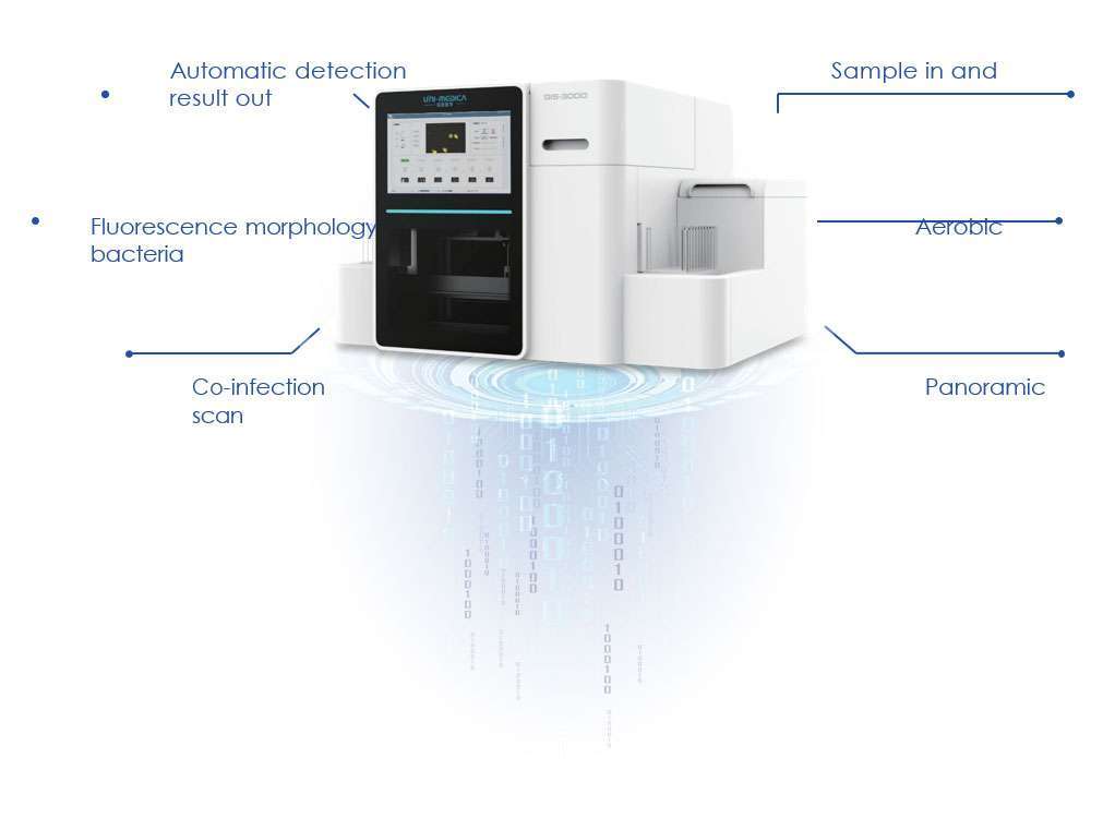
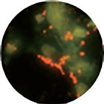
Automatic Gynecological Inflammation Intelligent Fluorescence Analysis System


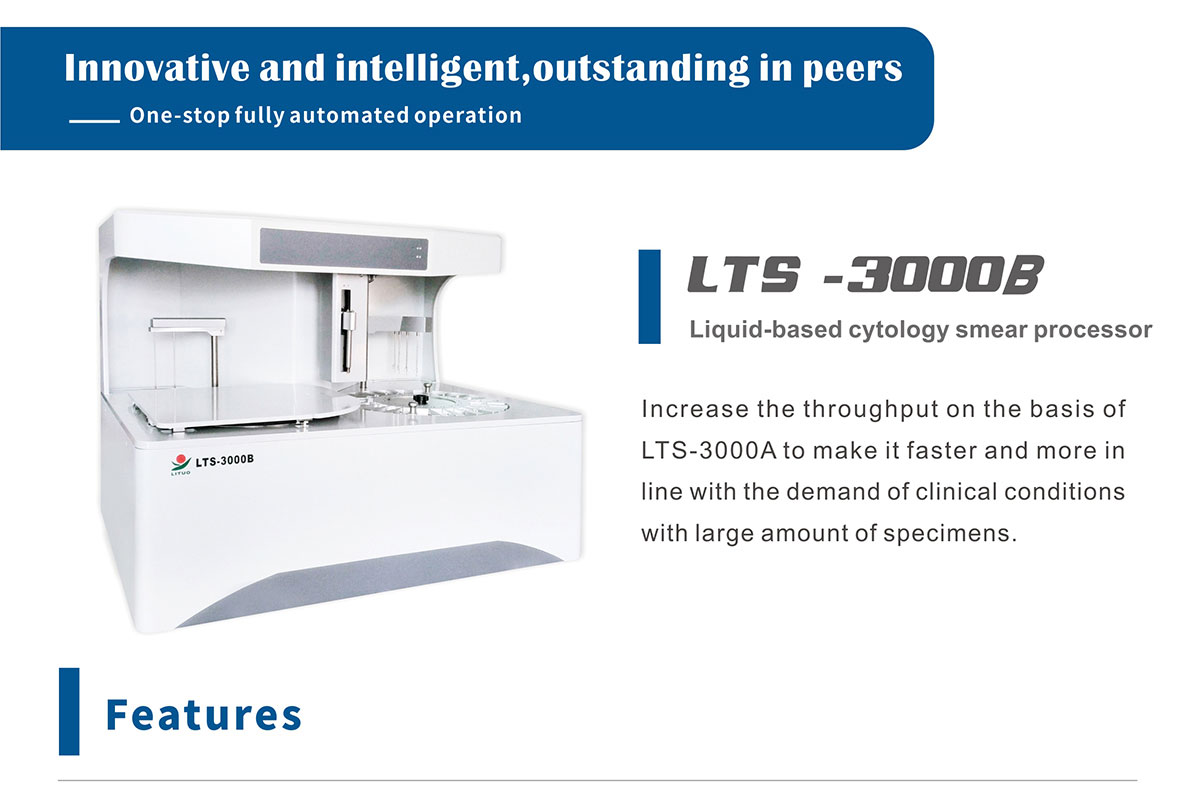
Pap Smear Cervical Screening LBC Automation