Women’s Health Vaginitis - Gynecological Inflammation Testing System
Artificial Intelligence Based Report
Gynecological inflammation intelligent
fluorescence analysis system
IntroductionofVaginitis
Vaginitis is the inflammation of the vagina, often caused by infections, bacteria, or yeast. It can lead to discomfort, itching, and unusual discharge, requiring prompt medical attention for proper diagnosis and treatment.
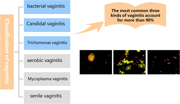
Classification of vaginitis
Single infection and mixed infection
More than 90% of vaginitis can be summarized as:
Bacterial vaginitis( 40%-50%)
Candidal vaginitis( 30%~40%)
Trichomonas vaginitis(10%-20%)1
40.5% of vaginitis is caused by 2 or more kinds of microorganisms!
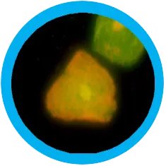
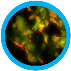
Multiplex Fluorescence Staining Technology
The multiple fluorescent staining technology has the dual advantages of fluorescence and morphology. It can stain Epithelial cells, Leukocytes, Lactobacilli, Clue cells, Candida vaginitis and Trichomonas vaginitis at the same time. It can not only detect vaginal related pathogen infection, but also evaluate whether the vaginal microecology and vaginal cleanliness are normal, providing help for rapid and accurate clinical diagnosis and treatment.
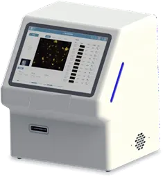
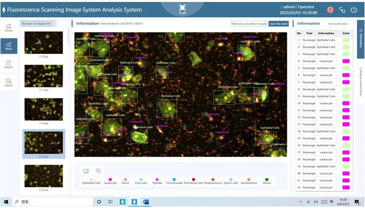
Automatic gynecological inflammation intelligent fluorescence analysis system Gis-1000
AI recognition, one-click report
Fast, scan 1 sample in 35 seconds
With blue and violet fluorescent modules, one-key automatic switching
Reports can be automatically adjusted for more flexibility
Automatic alarm function
Throughput: 1 sample
Gis1000
Automatic scanning and reading
Fully Automated Solution
Sample manufacture, staining, scanning and reading in one
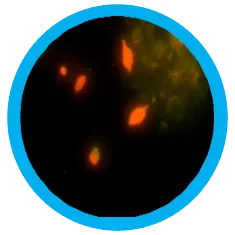
The multiple fluorescent staining technology has the dual advantages of fluorescence and morphology (Automated florescent Microscopy). When observed under the same fluorescent light source, it can show different staining effects on cells, bacteria and fungi, and simultaneously detect BV (bacterial vaginitis), TV (Trichomonas vaginitis) ), and VVC (Candida vaginitis).
Epithelial cells / leukocytes
green fluorescence
Clue cells
green fluorescence
Candida albicans
green fluorescence
Trichomonas
green fluorescence
Automatic scanning and reading
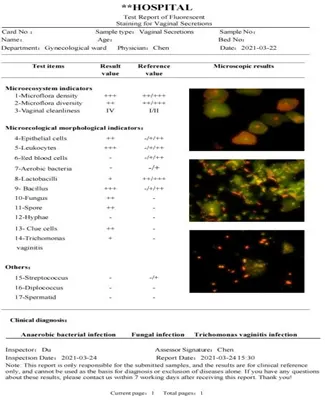
Clinical Report
Content of clinical report :
Clue cells:Interpret BV
Fungal hyphae/spores:Interpret VVC
Trichomonad:Interpret TV
Aerobes:Interpret AV
Number of leukocyte:Interpret cleanliness
Proportion of lactobacillus:Interpret vaginal microecology
Report results in the form of a combination of morphological pictures and text interpretation